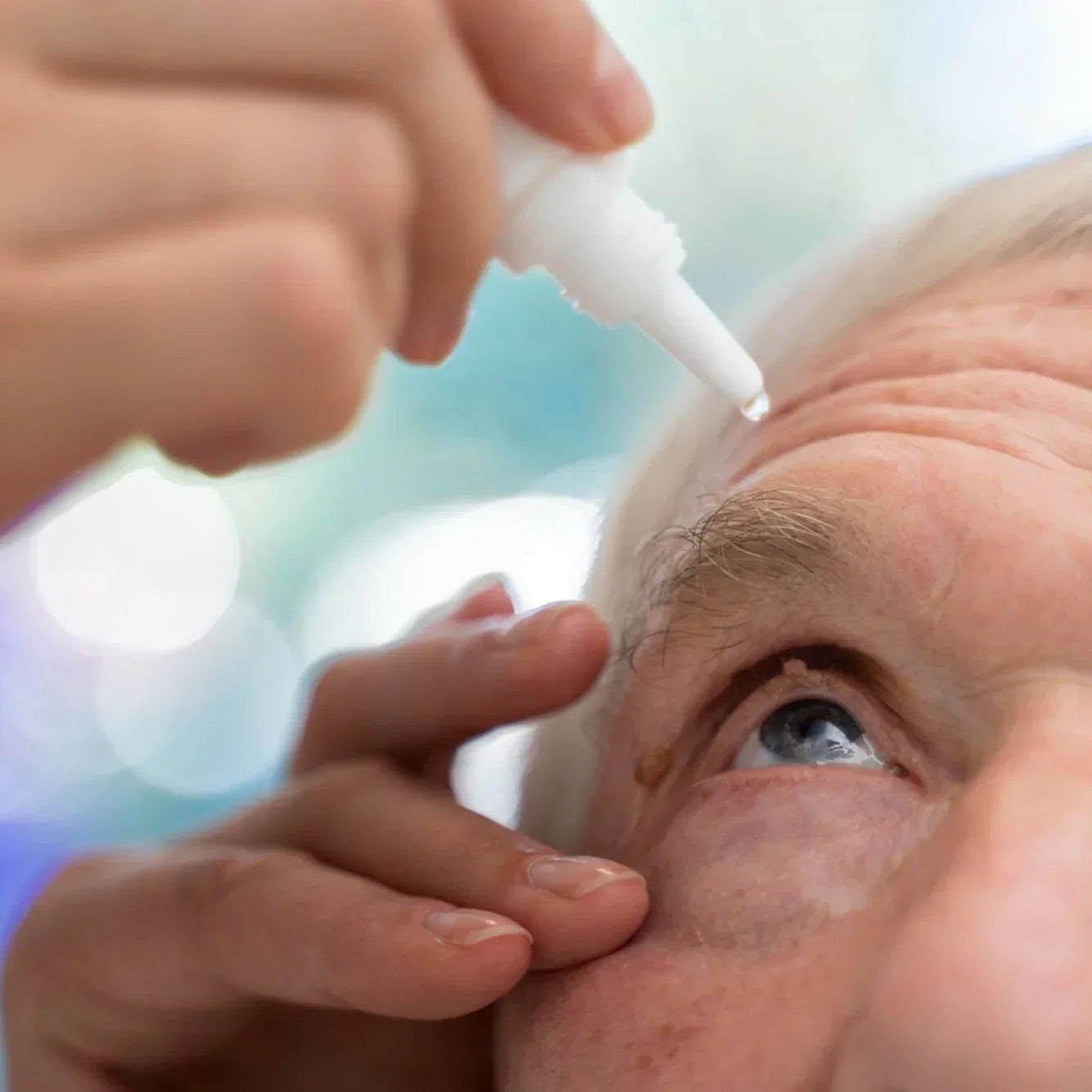
Glaucoma represents one of the leading causes of irreversible blindness worldwide, characterized fundamentally as a progressive optic neuropathy resulting in distinctive damage to the optic nerve head and corresponding visual field loss. The condition affects over 60 million people globally, with projections suggesting this number could reach 112 million by 20409. While intraocular pressure (IOP) remains the primary modifiable risk factor, substantial evidence indicates glaucoma's pathophysiology extends well beyond pressure mechanics to include neuroinflammation, oxidative stress, vascular dysfunction, and genetic factors. This comprehensive analysis explores the current understanding of glaucoma's complex mechanisms, the conventional and emerging treatment approaches, and distinguishes between evidence-based interventions and those requiring further validation. Understanding these multifaceted aspects is crucial for developing more effective therapeutic strategies to prevent irreversible vision loss in affected individuals.
Defining Glaucoma and Its Classification
Glaucoma encompasses a group of optic neuropathies characterized by the progressive loss of retinal ganglion cells (RGCs), resulting in characteristic optic nerve head damage and visual field defects20. The disease manifests in various forms, with primary open-angle glaucoma (POAG) being the most prevalent, accounting for approximately 75% of all cases worldwide9. POAG is characterized by RGC degeneration and optic nerve damage despite an anatomically open anterior chamber angle, typically developing insidiously and often remaining asymptomatic until significant vision loss occurs9. Other major classifications include angle-closure glaucoma, where physical obstruction of the drainage angle occurs, neovascular glaucoma (NVG) associated with abnormal blood vessel formation, and childhood glaucoma, a rare but serious condition that can lead to blindness if not diagnosed and treated promptly18.
The epidemiological burden of glaucoma continues to grow at an alarming rate, with approximately 20% of cases already at an incurable stage9. This increasing prevalence underscores the urgent need for improved early detection methods and more effective therapeutic interventions targeting the disease's underlying mechanisms. Child-onset glaucoma presents particular challenges, as it accompanies affected individuals throughout their lives and imposes significant burdens on families and healthcare systems18. Recent advances in molecular genetics and pathogenesis research have expanded our understanding of childhood glaucoma, potentially improving diagnosis and treatment approaches for this vulnerable population18.
Pathophysiological Mechanisms of Glaucoma
Intraocular Pressure and Mechanical Stress
Elevated intraocular pressure has traditionally been recognized as the primary risk factor for glaucoma development and progression20. The mechanical stress exerted on the optic nerve head, particularly at the lamina cribrosa (a mesh-like structure through which RGC axons pass to form the optic nerve), represents a critical mechanism of injury15. Sustained pressure can disrupt axonal transport systems, compromise blood flow to the optic nerve, and trigger a cascade of degenerative events ultimately leading to RGC death15. Recent research has revealed a fascinating paradox: under increased ocular pressure, retinal ganglion cells exhibit enhanced excitability, including improved response to light, even in cells with substantial dendritic pruning15. This adaptation apparently stems from voltage-dependent mechanisms in the axon and may serve as a compensatory response to maintain visual signaling despite structural damage15.
Despite the clear importance of IOP in glaucoma pathogenesis, the condition can develop and progress even in individuals with normal pressure readings, indicating the involvement of additional mechanisms13. Furthermore, in some patients, glaucomatous progression continues despite successful IOP reduction, suggesting that pressure-independent pathways contribute significantly to the disease process13. This clinical observation has driven extensive research into alternative pathogenic mechanisms and potential therapeutic targets beyond pressure control. Understanding these complex interactions between pressure-dependent and pressure-independent mechanisms is essential for developing more comprehensive treatment strategies that address the multifaceted nature of glaucomatous damage.
Trans-Lamina Cribrosa Pressure Gradient and Glymphatic System
An emerging concept in glaucoma pathophysiology involves the pressure differential across the lamina cribrosa19. Recent research has highlighted decreased intracranial pressure as a potential risk factor for glaucoma, as it creates an abnormally high trans-lamina cribrosa pressure difference19. This pressure gradient could disrupt normal fluid dynamics around the optic nerve, potentially affecting nutrient delivery and waste removal from this critical region. The discovery of a paravascular transport system in the eye, analogous to the brain's "glymphatic system," has provided new insights into potential mechanisms of glaucomatous damage19. This functional waste clearance pathway promotes elimination of interstitial solutes, including β-amyloid, along paravascular channels19.
The glymphatic hypothesis suggests that restriction of normal fluid flow at the level of the lamina cribrosa due to abnormal pressure gradients could impair the clearance of neurotoxic substances from the retina and optic nerve19. This mechanism is particularly compelling given that β-amyloid accumulation has been reported in glaucomatous models and is known to cause RGC death19. The inability to clear these toxic substances could contribute to chronic neurodegeneration even in the absence of significantly elevated IOP. This framework offers a novel perspective on how pressure dynamics might contribute to neurodegenerative changes in glaucoma and suggests potential therapeutic approaches aimed at enhancing glymphatic clearance mechanisms.
Neuroinflammation and Immune Responses
Chronic inflammation and immune-mediated damage constitute significant pathophysiological mechanisms in glaucoma development and progression12. Post-mortem studies of glaucomatous tissues have consistently demonstrated glial cell activation, with characteristic morphological and molecular changes in astrocytes, microglia, and to some extent, Müller cells12. These activated glial cells contribute to neuroinflammation through various pathways, including the release of pro-inflammatory cytokines, chemokines, and reactive oxygen species. Activated microglia typically adopt an amoeboid shape with increased expression of markers such as CD45 or HLA-DR, while astrocytes become hypertrophied with enhanced glial fibrillary acidic protein labeling12.
The inflammatory process in glaucoma appears to involve activation of danger-associated molecular patterns (DAMPs) and the classical complement cascade, creating an environment conducive to RGC damage12. Prolonged inflammatory responses lead to hypersecretion of inflammatory mediators and infiltration of inflammatory cells into affected tissues, potentially exacerbating the effects of increased IOP and ischemia9. Changes to extracellular matrix proteins like collagen, galectin, and tenascin-C suggest that glial cells influence structural alterations in the optic nerve head that may further compromise axonal integrity and function12. This chronic inflammatory state creates a self-perpetuating cycle that can accelerate disease progression independently of IOP elevation, explaining why some patients continue to deteriorate despite adequate pressure control.
Oxidative Stress and Excitotoxicity
Oxidative stress plays a pivotal role in RGC death in glaucoma, contributing significantly to disease progression through multiple pathways13. The retina, with its high oxygen consumption and exposure to light, is particularly vulnerable to oxidative damage from reactive oxygen species (ROS). Under glaucomatous conditions, impaired blood flow to the optic nerve head creates ischemic conditions that promote ROS generation, leading to damage of cellular components including DNA, proteins, and lipids13. This oxidative environment ultimately triggers mitochondrial dysfunction and activates apoptotic pathways in RGCs. The cumulative effects of chronic oxidative stress can overwhelm cellular antioxidant defense mechanisms, creating a state of redox imbalance that contributes to progressive neuronal loss.
Excitotoxicity represents another significant mechanism contributing to RGC degeneration in glaucoma13. This process typically involves excessive stimulation of glutamate receptors, particularly NMDA receptors, leading to calcium overload, mitochondrial dysfunction, and eventual neuronal death. Under glaucomatous conditions, glutamate can accumulate in the extracellular space due to impaired clearance mechanisms or increased release from damaged cells. The resulting overstimulation of glutamate receptors initiates a cascade of intracellular events that culminate in RGC apoptosis. These mechanisms of oxidative stress and excitotoxicity appear closely interrelated, with each potentially amplifying the other in a detrimental cycle that accelerates glaucomatous damage independent of IOP.
Vascular Factors and Ischemia
Circulatory disorders and ischemic events contribute substantially to glaucoma pathogenesis, particularly in certain forms of the disease9. Impaired blood flow to the optic nerve head can result from direct compression of blood vessels by elevated IOP or from systemic vascular dysregulation. This reduced perfusion creates a hypoxic environment that triggers a cascade of pathological events, including oxidative stress, excitotoxicity, and inflammatory responses. In neovascular glaucoma (NVG), which often develops secondary to proliferative diabetic retinopathy, ischemia-induced upregulation of vascular endothelial growth factor (VEGF) promotes the formation of abnormal blood vessels over the iris and anterior chamber angle1.
These newly formed vessels, along with accompanying fibrovascular tissue, can obstruct aqueous humor outflow, leading to severe IOP elevation and rapid vision loss if left untreated1. The increasing prevalence of diabetes worldwide has contributed to a rising incidence of NVG, making this particularly aggressive form of glaucoma an important public health concern1. The complex interplay between systemic vascular conditions and ocular pathology in NVG highlights the importance of comprehensive management approaches that address both ocular and systemic factors. Early diagnosis and aggressive treatment, including panretinal photocoagulation, anti-VEGF injections, and appropriate control of blood glucose levels in diabetic patients, are essential for halting the neovascularization process and preserving vision1.
Genetic Factors and Molecular Mechanisms
Genetic predisposition plays a significant role in glaucoma susceptibility and progression, with numerous genetic loci identified as contributing to disease risk9. Primary open-angle glaucoma often demonstrates complex inheritance patterns, involving interactions between multiple genes and environmental factors. Recent research has highlighted SVEP1 as a human coronary artery disease risk locus with causal links to both cardiometabolic disease and glaucoma, suggesting potential shared pathogenic mechanisms3. This gene has been identified as the first known physiologic ligand for PEAR1, a critical receptor governing platelet reactivity, with implications for understanding the vascular components of glaucoma pathogenesis3.
At the molecular level, glaucoma involves complex interactions between various signaling pathways and structural components12. Elevated IOP can induce changes in extracellular matrix proteins that contribute to structural alterations in the optic nerve head and trabecular meshwork12. These changes affect aqueous humor dynamics and potentially exacerbate disease progression through mechanical effects on RGC axons. The trabecular meshwork, responsible for draining aqueous humor from the anterior chamber, undergoes significant remodeling in glaucoma, contributing to outflow resistance and IOP elevation9. Understanding these molecular mechanisms provides valuable insights for developing targeted therapeutic approaches that address specific pathogenic pathways rather than simply lowering IOP.
Evidence-Based Treatment Approaches
Intraocular Pressure Reduction Strategies
Reducing intraocular pressure remains the cornerstone of glaucoma management, as it currently represents the only modifiable risk factor with proven efficacy in slowing disease progression20. Several well-established approaches exist for lowering IOP, including pharmacological agents, laser treatments, and surgical interventions. Topical medications constitute the first-line treatment for most glaucoma patients, with prostaglandin analogs generally preferred as initial therapy due to their superior IOP-lowering efficacy, once-daily dosing schedule, and relatively favorable side effect profile2. Other medication classes include beta-blockers, alpha-2 agonists, carbonic anhydrase inhibitors, and cholinergic agents, which may be used alone or in combination depending on individual patient needs and response.
Laser therapies, particularly selective laser trabeculoplasty (SLT), have gained prominence as effective interventions for IOP reduction2. Evidence indicates that SLT remains effective even in patients previously treated with prostaglandin analogs, providing an additional treatment option for those with inadequate IOP control on medical therapy or issues with medication adherence2. When target IOP cannot be achieved with less invasive methods, surgical procedures such as trabeculectomy may become necessary20. These filtering surgeries create an alternative pathway for aqueous humor outflow but require careful management of the wound healing process to prevent excessive scarring that could lead to filtration failure20. Anti-fibrotic agents such as mitomycin C and 5-fluorouracil are commonly employed during surgery to modulate the scarring process, though long-term failure rates remain significant due to the complexity of fibrotic mechanisms and limitations of current anti-scarring strategies20.
Comprehensive Management of Neovascular Glaucoma
Neovascular glaucoma, particularly when secondary to proliferative diabetic retinopathy, requires a multifaceted treatment approach addressing both the underlying ischemia and the resulting neovascularization1. Panretinal photocoagulation (PRP) serves as a fundamental treatment for the underlying retinal ischemia, reducing the production of angiogenic factors like VEGF by destroying oxygen-deprived retinal tissue1. Anti-VEGF injections have emerged as a valuable adjunctive therapy, directly targeting the key mediator of neovascularization and often producing rapid regression of iris and angle neovascularization1. These injections can effectively lower IOP and potentially delay or prevent the need for more invasive surgical interventions in many cases.
In addition to these specific interventions, conventional anti-glaucoma medications play an important role in managing IOP while more definitive treatments take effect1. For patients with diabetes, proper control of blood glucose levels is essential for addressing the underlying cause of retinal ischemia and preventing disease progression1. Early diagnosis and aggressive treatment remain crucial in halting the neovascularization process and preserving vision in these challenging cases, as NVG can progress rapidly and cause severe visual impairment if left untreated1. The management of NVG exemplifies the importance of understanding specific disease mechanisms in developing effective treatment strategies that address not only elevated IOP but also the underlying pathogenic processes driving disease progression.
Neuroprotective Approaches: Evidence and Limitations
Given that glaucoma progression can continue despite adequate IOP control in some patients, there is growing interest in neuroprotective strategies that directly target RGC survival independently of pressure mechanics13. These approaches aim to interrupt the various pathways leading to RGC death, including oxidative stress, excitotoxicity, and neuroinflammation. Several natural products have shown promise in protecting RGCs from glaucomatous damage through multiple mechanisms13. Ginkgo biloba, Lycium barbarum, Diospyros kaki, Tripterygium wilfordii, saffron, curcumin, caffeine, anthocyanin, coenzyme Q10, and vitamins B3 and D have demonstrated neuroprotective effects primarily through antioxidant, anti-inflammatory, and anti-apoptotic actions13.
Some natural compounds appear capable of directly reducing IOP through various mechanisms13. Baicalein, forskolin, marijuana, ginsenoside, resveratrol, and hesperidin have been reported to lower IOP in experimental models and limited clinical studies13. Marijuana's potential role in glaucoma treatment has generated particular interest, as it may have beneficial effects based on its mechanism of action in the eye7. Despite these promising findings, most neuroprotective approaches remain inadequately validated by large-scale clinical trials, and their long-term efficacy and safety profiles require further investigation13. The translation of experimental neuroprotective strategies into clinical practice has generally been challenging, with numerous agents showing promise in laboratory studies but failing to demonstrate comparable benefits in human trials. This discrepancy highlights the complexity of glaucomatous neurodegeneration and the need for better models that more accurately reflect human disease processes.
Emerging Therapeutic Approaches and Future Directions
Targeting Molecular Pathways and Inflammation
Advanced understanding of the molecular mechanisms underlying glaucoma has identified several potential therapeutic targets beyond conventional IOP reduction1213. Anti-inflammatory and immunomodulatory approaches aim to interrupt the chronic neuroinflammation that contributes to RGC death regardless of pressure status13. Cyclosporin (CyA), an immunosuppressive agent, has shown efficacy in various ocular inflammatory conditions and might potentially benefit glaucoma patients by attenuating inflammatory responses that contribute to RGC damage8. The application of 0.1% CyA eye drops represents a specific therapeutic approach that has received orphan drug designation from the European Commission for certain ocular conditions, though its application in glaucoma management requires further investigation8.
Manipulating specific signaling pathways involved in glaucoma pathogenesis offers another promising avenue for intervention3. Recent research has illuminated the interactions between SVEP1, PEAR1, and the Ang/Tie pathway, with potential therapeutic implications for both glaucoma and related systemic conditions3. Targeting these molecular interactions could potentially address underlying disease mechanisms rather than simply treating symptoms or risk factors. Additionally, approaches targeting the extracellular matrix and structural remodeling in the trabecular meshwork and optic nerve head might help prevent the progressive changes that contribute to IOP elevation and axonal damage in glaucoma12. While these molecular approaches remain largely experimental, they represent an important frontier in glaucoma research that could eventually transform treatment paradigms.
Artificial Intelligence and Advanced Diagnostics
The application of artificial intelligence (AI) in glaucoma diagnosis and management represents a rapidly evolving field with significant potential to improve clinical outcomes5. Deep convolutional neural networks have demonstrated impressive capabilities in analyzing fundus retinal images for glaucoma detection, with some models achieving training set accuracy rates approaching 98%5. These AI systems can identify subtle patterns and changes that might escape human detection, potentially enabling earlier diagnosis and intervention before significant vision loss occurs. The integration of AI-based algorithms with other diagnostic technologies could potentially revolutionize glaucoma screening and monitoring, particularly in resource-limited settings where specialist care may be unavailable.
Models such as AlexNet and LeNet, when enhanced with Batch Normalization layers to improve convergence speed and processing efficiency, have shown particular promise in glaucoma image analysis5. These technological advances complement the growing understanding of glaucoma's biological mechanisms and could eventually enable more personalized treatment approaches based on individual risk profiles and disease characteristics. The development of AI-based risk calculators represents another important application, potentially allowing for evidence-based care of ocular hypertension and glaucoma patients through more accurate prediction of disease onset and progression16. While these technologies continue to evolve and require further validation in diverse clinical settings, they exemplify how technological innovation can complement biological insights to improve glaucoma management.
Novel Drug Delivery Systems and Combination Therapies
Challenges with treatment adherence and ocular drug bioavailability have stimulated interest in advanced drug delivery systems for glaucoma management813. Sustained-release formulations, implantable devices, and innovative vehicle technologies aim to overcome the limitations of conventional eye drops, which often suffer from poor ocular penetration and require frequent administration. These delivery systems could potentially improve treatment efficacy while reducing the burden on patients and enhancing adherence to therapy regimens. Additionally, approaches that target multiple pathogenic mechanisms simultaneously through combination therapies may prove more effective than single-mechanism interventions, particularly given the complex and multifactorial nature of glaucomatous damage20.
Recent research suggests that a better approach to manage post-surgical fibrosis may involve targeting multiple points in the scarring process, thereby increasing the inhibitory potential against excessive tissue remodeling20. This principle likely applies to glaucoma management more broadly, where addressing multiple pathogenic mechanisms simultaneously may produce synergistic benefits. For instance, combining IOP-lowering strategies with neuroprotective agents, anti-inflammatory treatments, and approaches to enhance waste clearance through the glymphatic system could potentially provide more comprehensive protection against progressive RGC loss1319. While such multimodal approaches increase treatment complexity and potentially introduce new challenges related to drug interactions and combined toxicities, they may ultimately prove necessary to adequately address the multifaceted nature of glaucomatous damage.
Conclusion
Glaucoma represents a complex neurodegenerative disorder with multiple pathophysiological mechanisms extending well beyond elevated intraocular pressure. Current evidence highlights the importance of mechanical stress at the lamina cribrosa, neuroinflammation, oxidative damage, vascular dysregulation, genetic factors, and potentially impaired waste clearance through the glymphatic system in disease development and progression. While IOP reduction remains the cornerstone of treatment with the strongest evidence base, the limitations of pressure-centric approaches have become increasingly apparent as our understanding of glaucoma's complexity has evolved. Many patients continue to experience disease progression despite apparently adequate IOP control, underscoring the need for complementary therapeutic strategies targeting pressure-independent mechanisms of damage.
The therapeutic landscape for glaucoma continues to evolve, with well-established approaches like medications, laser therapy, and surgery being supplemented by emerging strategies targeting neuroprotection, inflammation modulation, and specific molecular pathways. Natural products with antioxidant and anti-inflammatory properties show promise as potential adjunctive therapies, though most require further validation through rigorous clinical trials. Advanced technologies, particularly artificial intelligence and innovative drug delivery systems, offer exciting possibilities for improving diagnosis, monitoring, and treatment of this challenging condition. As our understanding of glaucoma mechanisms continues to deepen, a more comprehensive approach to management will likely emerge, combining traditional IOP-lowering strategies with novel interventions targeting the fundamental neurodegenerative processes that ultimately result in vision loss. Such multimodal approaches may eventually prove more effective in preserving vision for the millions of individuals affected by glaucoma worldwide.
Citations:
- https://www.ncbi.nlm.nih.gov/pmc/articles/PMC9900735/
- https://pubmed.ncbi.nlm.nih.gov/35962295/
- https://pubmed.ncbi.nlm.nih.gov/39847464/
- https://www.ncbi.nlm.nih.gov/pmc/articles/PMC9862076/
- https://www.semanticscholar.org/paper/a1192fee80c68be5f632aa8582ff96c7381faf2f
- https://pubmed.ncbi.nlm.nih.gov/40029267/
- https://www.semanticscholar.org/paper/dee1491f29346ef0b540ee4c18c2032632e6d4fb
- https://www.ncbi.nlm.nih.gov/pmc/articles/PMC6616155/
- https://www.semanticscholar.org/paper/217b1d3b43f991169c862123bee4b7c431ab9bec
- https://pubmed.ncbi.nlm.nih.gov/25663482/
- https://www.semanticscholar.org/paper/8fb7a69c4a007db1002693783f99ce1f3569e03c
- https://www.ncbi.nlm.nih.gov/pmc/articles/PMC11346413/
- https://www.ncbi.nlm.nih.gov/pmc/articles/PMC8840399/
- https://www.semanticscholar.org/paper/e425d671b3b435b26bbb90c7fbdd7c6111100933
- https://www.ncbi.nlm.nih.gov/pmc/articles/PMC5877940/
- https://www.semanticscholar.org/paper/b0b35724a221bdcced23b613563ab8760904ae9c
- https://pubmed.ncbi.nlm.nih.gov/18260297/
- https://pubmed.ncbi.nlm.nih.gov/38706086/
- https://pubmed.ncbi.nlm.nih.gov/28129671/
- https://www.ncbi.nlm.nih.gov/pmc/articles/PMC10305204/
- https://www.semanticscholar.org/paper/b35770e8e8f5cfa7bc193243ec6a09809504edf3
- https://pubmed.ncbi.nlm.nih.gov/18260296/
- https://pubmed.ncbi.nlm.nih.gov/35037367/
- https://www.ncbi.nlm.nih.gov/pmc/articles/PMC4228301/
- https://www.ncbi.nlm.nih.gov/pmc/articles/PMC11228121/
- https://www.ncbi.nlm.nih.gov/pmc/articles/PMC11591249/
- https://pubmed.ncbi.nlm.nih.gov/39760712/
- https://pubmed.ncbi.nlm.nih.gov/39977697/
- https://pubmed.ncbi.nlm.nih.gov/39992791/
- https://pubmed.ncbi.nlm.nih.gov/29456252/


