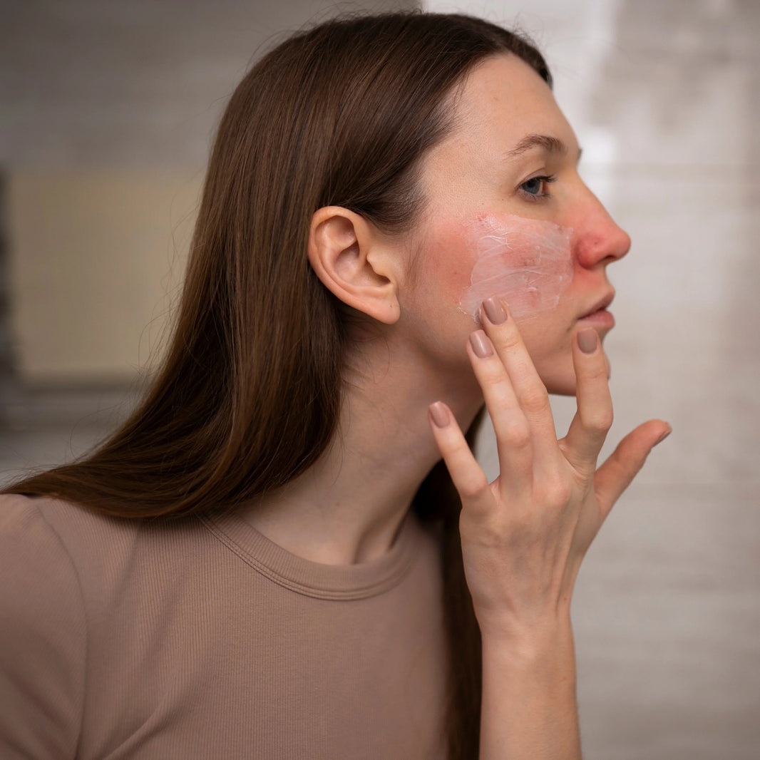
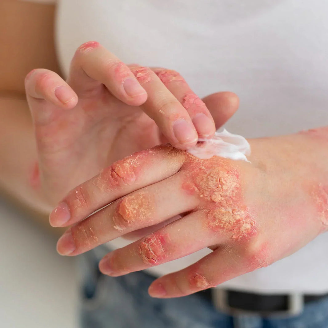
Dermatitis represents a complex family of inflammatory skin conditions characterized by distinctive pathophysiological mechanisms and clinical manifestations. This report provides a comprehensive analysis of the various forms of dermatitis, exploring their underlying mechanisms, molecular pathways, therapeutic targets, and evidence-based treatment approaches. Understanding the intricate interplay between genetic predisposition, environmental triggers, and immunological dysregulation offers valuable insights into effective management strategies for this prevalent skin disorder that significantly impacts patients' quality of life.
The Spectrum of Dermatitis Conditions
Dermatitis encompasses several distinct but related inflammatory skin disorders, with atopic dermatitis (AD) being the most prevalent and extensively studied form. AD is a chronic, recurrent inflammatory skin disorder manifesting as eczematous lesions accompanied by intense pruritus (itching), affecting more than 10% of children worldwide1112. This condition frequently develops before the onset of other atopic diseases such as allergic rhinitis or asthma, establishing itself as a significant entry point in the atopic march11. The disease burden extends beyond physical symptoms, profoundly affecting patients' psychological wellbeing and quality of life, particularly when the condition becomes chronic.
Contact dermatitis represents another common form, categorized into allergic contact dermatitis (ACD) and irritant contact dermatitis. ACD manifests as a type IV delayed-type hypersensitivity reaction triggered by allergen exposure in previously sensitized individuals through the activation of allergen-specific T cells17. In its acute phase, ACD presents with eczematous dermatitis characterized by erythema, edema, vesicles, scaling, and intense itching. Non-eczematous clinical variants also exist, including lichenoid, bullous, and lymphomatous forms17. If the culprit allergen remains unidentified or exposure continues, the condition commonly progresses to lichenification in the chronic phase.
Other dermatitis variants include radiation-induced dermatitis, a common complication in cancer patients undergoing radiotherapy, particularly those with breast, head and neck, anal, and vulvar cancers7. Stasis dermatitis, associated with venous insufficiency, and arsenic-related dermatitis resulting from environmental toxin exposure represent additional specialized forms with distinct pathophysiological mechanisms59. Each variant exhibits unique clinical and histopathological features while sharing certain fundamental inflammatory pathways.
Fundamental Pathophysiology of Dermatitis
The pathogenesis of dermatitis revolves around three interconnected pillars: epidermal barrier dysfunction, immune system dysregulation, and neuroinflammatory processes leading to pruritus13. While specific mechanisms vary across different forms, these core pathophysiological elements remain consistent.
Epidermal barrier impairment represents a critical inciting factor in dermatitis development. In AD, genetic predisposition to barrier dysfunction, often linked to filaggrin gene mutations, creates vulnerability that environmental factors can exploit11. The compromised barrier allows penetration of allergens, irritants, and pathogens, triggering inflammatory cascades. This initial barrier dysfunction initiates a vicious cycle where inflammation further deteriorates barrier integrity, perpetuating the disease process.
Immunological imbalance constitutes the second fundamental pillar of dermatitis pathophysiology. In lesional skin of AD, various innate immune cells, including Th2 cells, type 2 innate lymphoid cells (ILC2s), and basophils, produce signature Th2 cytokines (interleukin-4, IL-5, IL-13, IL-31)13. Epidermal keratinocytes release alarmins such as thymic stromal lymphopoietin (TSLP), IL-25, and IL-33, which amplify type 2 inflammation. As the condition progresses to chronicity, the immunological landscape evolves to include Th22 and Th17 cells, creating a more complex inflammatory milieu13.
Atopic Dermatitis: Molecular Mechanisms and Pathways
The pathophysiology of AD involves intricate interactions between genetic predisposition, environmental factors, immune dysregulation, and barrier dysfunction. Recent advances have illuminated the complex molecular orchestra orchestrating this condition.
Langerhans cells (LCs), specialized dendritic cells residing in the epidermis, emerge as central players in AD pathophysiology15. As professional antigen-presenting cells, LCs detect environmental antigens and allergens, subsequently shaping adaptive immune responses through T-cell polarization. Their sentinel function extends to monitoring the skin microbiome and influencing immune tolerance versus reactivity decisions. Beyond immunological functions, LCs participate in maintaining skin barrier integrity by modulating tight junctions, and they serve as crucial mediators in neuro-immune crosstalk within the skin15.
The cytokine profile in AD exhibits dynamic changes throughout disease progression. In acute lesions, the Th2 signature predominates, with elevated IL-4, IL-13, and IL-31 levels. The chronic phase witnesses a broader cytokine landscape with additional contributions from Th22 (IL-22) and Th17 (IL-17) pathways13. This evolving cytokine milieu directly impairs skin barrier function, with IL-4, IL-13, and IL-22 suppressing filaggrin expression, thereby exacerbating barrier dysfunction13. The resulting molecular environment creates self-perpetuating inflammation cycles that maintain disease chronicity.
Circadian rhythm disruption represents an emerging contributor to AD pathophysiology. Research indicates that circadian rhythm influences cutaneous blood flow and skin barrier properties, including transepidermal water loss and capacitance8. Aberrations in these circadian patterns may exacerbate barrier dysfunction and immune dysregulation, suggesting chronotherapy as a potential targeted approach to improve treatment outcomes in AD patients8.
Contact Dermatitis: Immunological Mechanisms
Contact dermatitis, particularly allergic contact dermatitis (ACD), operates through distinct immunological mechanisms from atopic dermatitis. ACD represents a classic example of type IV delayed-type hypersensitivity reaction occurring in two phases: sensitization and elicitation17.
The sensitization phase begins when haptens (small molecular weight chemicals) penetrate the skin and bind to carrier proteins, forming complete antigens. Dendritic cells process these antigens and migrate to regional lymph nodes, where they present the processed antigens to naïve T cells. This interaction leads to the development of memory T cells specific to the allergen, establishing immunological memory without causing clinical symptoms17.
The elicitation phase occurs upon re-exposure to the same allergen. The previously sensitized individual mounts a rapid immune response where memory T cells recognize the allergen, proliferate, and release proinflammatory cytokines. This orchestrated immune reaction manifests clinically as the characteristic eczematous skin lesions 24-72 hours after allergen exposure17.
Common allergens implicated in ACD include metals (particularly nickel), fragrance mix compounds, isothiazolinones, and para-phenylenediamine. These allergens represent both occupational and non-occupational exposures, with ACD accounting for approximately 90% of occupational skin disorders alongside irritant contact dermatitis17.
Molecular Targets for Therapeutic Intervention
The evolving understanding of dermatitis pathophysiology has revealed numerous molecular targets for therapeutic intervention. These targets primarily involve key mediators in inflammatory cascades and barrier function regulation.
Interleukins represent primary targets in AD management. IL-4 and IL-13, signature Th2 cytokines, promote IgE production, barrier dysfunction, and itch sensitization13. Dupilumab, a monoclonal antibody targeting the shared IL-4 receptor alpha subunit, effectively blocks both IL-4 and IL-13 signaling, demonstrating remarkable efficacy in moderate-to-severe AD13. IL-31, known as the "itch cytokine," directly activates sensory neurons and induces pruritus. Nemolizumab, an IL-31 receptor antagonist, has shown promise in reducing AD-associated itch13. IL-22, produced by Th22 cells, impairs keratinocyte differentiation and barrier function. Clinical trials with fezakinumab, targeting IL-22, have demonstrated efficacy particularly in severe AD cases13.
Janus kinase (JAK) inhibitors represent an emerging class of small molecule therapies targeting intracellular signaling pathways downstream of multiple cytokine receptors. By inhibiting JAK-STAT signaling, these molecules simultaneously attenuate multiple inflammatory pathways. Baricitinib, upadacitinib, and abrocitinib belong to this therapeutic class, offering potential advantages through broader immunomodulation compared to biologics targeting single cytokines1112.
Enzymes involved in inflammatory mediator synthesis present additional therapeutic targets. 5-lipoxygenase (5-LOX) and cyclooxygenase-2 (COX-2) are key enzymes in the arachidonic acid pathway, producing leukotrienes and prostaglandins that contribute to inflammation and pruritus in dermatitis14. Inhibitors of these enzymes may offer symptomatic relief by reducing proinflammatory lipid mediator production.
The skin barrier itself represents a fundamental therapeutic target. Strategies aimed at barrier repair and maintenance, including emollients and moisturizers containing ceramides, fatty acids, and cholesterol, help restore lipid composition and improve barrier function. This approach addresses a primary pathophysiological mechanism while potentially reducing the need for anti-inflammatory treatments10.
Evidence-Based Therapeutic Approaches
The therapeutic management of dermatitis follows a stepwise approach based on disease severity, chronicity, and specific subtype. Evidence-based guidelines emphasize the importance of individualized treatment strategies rather than rigid standardized protocols.
For atopic dermatitis, basic therapy centers on addressing barrier dysfunction through consistent application of hydrating and lubricating topical treatments, combined with avoidance of specific and non-specific trigger factors10. This foundational approach remains essential regardless of disease severity or concomitant treatments.
Topical anti-inflammatory agents constitute first-line therapy for mild-to-moderate AD. Topical corticosteroids remain the mainstay treatment, offering rapid and effective inflammation control. Calcineurin inhibitors, including tacrolimus and pimecrolimus, provide steroid-sparing alternatives particularly valuable for sensitive skin areas (face, genitals, skin folds) and long-term maintenance therapy10. Topical phosphodiesterase inhibitors represent an emerging alternative, though their availability varies by region10.
For moderate-to-severe AD unresponsive to topical therapies, systemic treatment options include conventional immunosuppressants and newer targeted biologics. Dupilumab, targeting IL-4/IL-13 signaling, has revolutionized AD management, demonstrating significant efficacy in reducing disease severity, pruritus, and improving quality of life13. This biologic has established a favorable safety profile compared to conventional immunosuppressants like cyclosporine or methotrexate.
Adjunctive phototherapy, particularly narrowband UVB, offers another evidence-based approach for moderate AD. This treatment modality exerts immunomodulatory effects, reducing inflammatory cell infiltration and cytokine production10. Proper patient selection and monitoring are essential to balance efficacy with potential long-term risks.
In allergic contact dermatitis, the primary and most effective intervention involves identifying and eliminating the offending allergen. Patch testing represents the diagnostic gold standard to identify culprit allergens17. Once identified, allergen avoidance prevents disease recurrence. For acute flares, topical corticosteroids provide symptomatic relief and inflammation control. Systemic corticosteroids may be necessary for severe or widespread reactions17.
Emerging and Less-Proven Therapeutic Approaches
The therapeutic landscape for dermatitis continues to evolve, with numerous emerging approaches showing preliminary promise but requiring further validation.
Several plant-derived compounds demonstrate potential anti-inflammatory properties relevant to dermatitis. Spirodela polyrhiza extract has shown efficacy in ameliorating skin lesions and reducing inflammatory cytokines (TNF-α, IFN-γ, IL-6, MCP-1) in experimental models2. Active compounds from this plant, particularly luteolin and apigenin, exhibited strong binding affinity for TNF and IL-6, suggesting specific molecular targets underlying their effects2. Similarly, Boesenbergia rotunda extract and its flavonoid pinostrobin demonstrated potent 5-LOX and COX-2 inhibitory activity, potentially addressing both inflammation and pruritus in atopic dermatitis14.
Targeting alarmins represents another emerging approach with mixed results. While IL-33, TSLP, and IL-25 play significant roles in AD pathophysiology by amplifying type 2 inflammation, clinical trials targeting these molecules have yielded inconsistent outcomes13. Despite promising results in murine models and correlation with disease severity in human patients, treatments targeting IL-33 specifically have not demonstrated sufficient efficacy in clinical trials13. This discrepancy highlights the complexity of translating preclinical findings to human disease.
Chronotherapy, tailoring treatment timing to align with circadian rhythm patterns, represents an innovative approach based on emerging understanding of circadian influences on skin barrier function and immune activity8. While conceptually promising, robust clinical evidence supporting this approach remains limited, necessitating further investigation to establish optimal implementation strategies and quantify clinical benefits.
Microbiome modulation strategies are also gaining attention. The skin microbiome dysbiosis, particularly Staphylococcus aureus colonization, contributes to AD pathophysiology. Approaches targeting microbiome restoration or inhibiting S. aureus virulence factors show theoretical promise14. However, translating these concepts into effective clinical interventions requires further development and validation.
Conclusion
Dermatitis represents a complex spectrum of inflammatory skin conditions with multifaceted pathophysiology involving barrier dysfunction, immune dysregulation, and neuroinflammatory mechanisms. Recent advances in understanding the molecular underpinnings of these conditions have revolutionized therapeutic approaches, moving from broad immunosuppression toward targeted interventions addressing specific disease mechanisms.
The therapeutic landscape continues to evolve rapidly, with biologics targeting key cytokines and small molecules inhibiting intracellular signaling pathways demonstrating promising efficacy in moderate-to-severe disease. However, basic therapy addressing barrier dysfunction remains fundamental across disease severity spectrum. Future directions include further refinement of targeted therapies, development of reliable biomarkers for endotyping to enable personalized medicine approaches, and exploration of combination strategies to address multiple disease aspects simultaneously.
While significant progress has been made, substantial challenges persist. The heterogeneity of dermatitis necessitates individualized approaches, and a subset of patients remains refractory to available treatments. Additionally, long-term safety profiles of newer therapies require continued surveillance. Collaborative efforts between clinicians, researchers, and patients will be essential to advance our understanding and optimize management strategies for this impactful group of skin disorders.
Citations:
- https://pubmed.ncbi.nlm.nih.gov/29063428/
- https://www.ncbi.nlm.nih.gov/pmc/articles/PMC10488168/
- https://www.ncbi.nlm.nih.gov/pmc/articles/PMC8959491/
- https://www.semanticscholar.org/paper/e40beb5258ef97ee2fc3bb3d9062194c0d65ec19
- https://pubmed.ncbi.nlm.nih.gov/28063094/
- https://www.ncbi.nlm.nih.gov/pmc/articles/PMC8605491/
- https://www.semanticscholar.org/paper/6d0e716b257529d95dd686d6d653b1f445f8865c
- https://pubmed.ncbi.nlm.nih.gov/29231268/
- https://www.ncbi.nlm.nih.gov/pmc/articles/PMC9171494/
- https://pubmed.ncbi.nlm.nih.gov/29676534/
- https://www.ncbi.nlm.nih.gov/pmc/articles/PMC10931328/
- https://www.ncbi.nlm.nih.gov/pmc/articles/PMC9820829/
- https://www.ncbi.nlm.nih.gov/pmc/articles/PMC10999685/
- https://www.ncbi.nlm.nih.gov/pmc/articles/PMC10812521/
- https://www.ncbi.nlm.nih.gov/pmc/articles/PMC11760705/
- https://pubmed.ncbi.nlm.nih.gov/38877357/
- https://www.ncbi.nlm.nih.gov/pmc/articles/PMC10239928/
- https://pubmed.ncbi.nlm.nih.gov/26905599/
- https://www.semanticscholar.org/paper/614800a79049e47fed73ace1cd3c5ad612294d43
- https://pubmed.ncbi.nlm.nih.gov/27129696/
- https://www.ncbi.nlm.nih.gov/pmc/articles/PMC9191759/
- https://www.semanticscholar.org/paper/636136fa236c199783366e9ff2a8e1ec855ada0a
- https://www.ncbi.nlm.nih.gov/pmc/articles/PMC8910412/
- https://pubmed.ncbi.nlm.nih.gov/37254967/
- https://pubmed.ncbi.nlm.nih.gov/37316462/
- https://pubmed.ncbi.nlm.nih.gov/30287314/
- https://www.ncbi.nlm.nih.gov/pmc/articles/PMC11084351/
- https://www.ncbi.nlm.nih.gov/pmc/articles/PMC10151557/
- https://pubmed.ncbi.nlm.nih.gov/33147660/
- https://pubmed.ncbi.nlm.nih.gov/35188464/
- https://pubmed.ncbi.nlm.nih.gov/39636193/
- https://www.ncbi.nlm.nih.gov/pmc/articles/PMC6109028/
- https://pubmed.ncbi.nlm.nih.gov/38724796/
- https://pubmed.ncbi.nlm.nih.gov/32579457/
- https://pubmed.ncbi.nlm.nih.gov/11140380/
- https://pubmed.ncbi.nlm.nih.gov/28008787/
- https://www.semanticscholar.org/paper/456aa84b3f27effe39c74b93b8b10b603c6e1b5c
- https://www.semanticscholar.org/paper/a86f4749a015f211565da680110a3cadc1a679e8
- https://pubmed.ncbi.nlm.nih.gov/38287887/
- https://www.ncbi.nlm.nih.gov/pmc/articles/PMC10095524/


